If you are a teen or young adult, and still have your wisdom teeth, you should probably have them evaluated before they begin to cause problems, such as pain, infection, or damage to other teeth. If you are experiencing complications from your third molars, now is the time to consider wisdom teeth removal. This extremely common procedure is an important step toward optimal oral health for many people.
Baltimore patients choose The Maryland Center for Oral Surgery and Dental Implants because we offer the expertise of surgeons, who remove wisdom teeth and administer anesthesia on a daily basis. In addition, we have a dedicated surgical facility with the most advanced equipment available for optimal patient safety and comfort.
What Are Wisdom Teeth?
Most people develop 32 permanent teeth over their lifetime. Typically, the last four teeth to erupt from the gums are the third molars, or “wisdom teeth.” These teeth normally emerge between the ages of 17 and 21, though they may emerge as early as age 11.
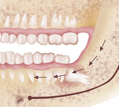
It is quite common for patients to require wisdom teeth removal since the jaws are rarely large enough to accommodate the third molars. As a result, these teeth can become “impacted”: embedded in gum tissue or bone beyond their normal eruption time.
The Maryland Center recommends wisdom teeth removal for those whom are reaching adulthood for several reasons:
As the teeth develop, the roots continue to grow longer, and the jawbone becomes denser. This increases the difficulty and complexity of removing the teeth.
The more established the wisdom teeth become, the more discomfort you may have following surgery.
After age 30, people are much more likely to experience the problems associated with impacted teeth.
Why Choose the Maryland Center for Oral Surgery Dental Implants for Wisdom Tooth Removal?
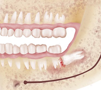
The main reason that you should consider extracting wisdom teeth is to avoid discomfort and oral health issues. These include:
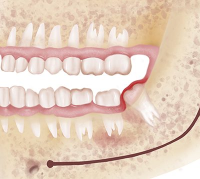
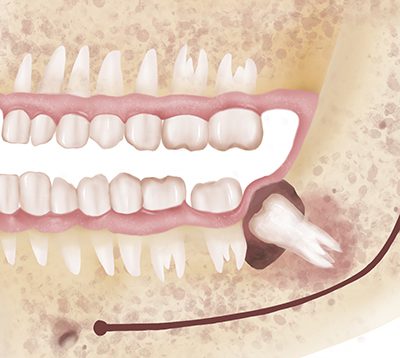
Wisdom teeth removal is more than just a healthy decision. It’s a way of preserving your smile. Impacted wisdom teeth can cause crowding, affect the alignment of your other teeth, and emerge at an incorrect angle from the gums. Extracting them before there’s a problem helps prevent these concerns.
During your initial consultation with one of our surgeons at the Maryland Center for Oral Surgery and Dental Implants, we will:
Conduct an examination and discuss the need for extracting your wisdom teeth.
Review your options for anesthesia, including local anesthesia, nitrous oxide analgesia (“laughing gas”), sedation, and general anesthesia. Our team wants to ensure that you are as comfortable as possible and completely pain-free during the surgery.
Explain what you can expect during and after your procedure. We will also give you instructions to prepare for the day of your surgery.
Our Oral Surgeons perform wisdom teeth removal in an environment that prioritizes safety. Doctors with years of experience in anesthesia techniques monitor your procedure and utilize top of the line surgical equipment. Your treatment plan will be tailored to your needs, which may vary based on the position of your wisdom teeth, whether they are erupted or impacted, and the position of the tooth roots.
After your procedure, you will go home with post-operative instructions, which we will also review with the person taking you home. Our website also provides this information. Click here for detailed post-surgical instructions.
Many patients manage discomfort with ibuprofen, but we will also provide a prescription for pain medication and possibly a prescription for an antibiotic. Within about 48 hours, your swelling should decrease, and you should be progressing well in your recovery. Throughout your recuperation, we encourage you to contact our team with any questions or concerns. It is of paramount concern to the surgeons and staff at the Maryland center for Oral Surgery and Dental Implants that you experience a comfortable and easy recovery.
For patients in the Baltimore area, there is no better place to get wisdom teeth removed than at Maryland Center for Oral Surgery and Dental Implants. With the latest in technology, the finest in staff, and the most complete facility around, the professionals at this facility can ensure the highest quality of care and comfort to everyone. Patients who are in need of wisdom teeth removal are encouraged to call (410) 628-1839 for a consultation today.
Our Surgeons Have Extensive Training and Education.
Please be aware that this is not a secure email network under HIPAA guidelines. Do not submit any personal or private information unless you are authorized and have voluntarily consented to do so. We are not liable for any HIPAA violations. Understand that if you email us, you are agreeing to the use of an unsecured method and understand that all replies will be sent in the same fashion, which you are hereby authorizing.
By checking this box you hereby agree to hold The Maryland Center for Oral Surgery and Dental Implants, including its doctors and affiliates, harmless from any hacking or any other unauthorized use of your personal information by outside parties. By checking this box, you also agree to receive email communication from The Maryland Center for Oral Surgery and Dental Implants.
This website is provided for information and education purposes only and is not intended to offer specific medical or surgical advice to anyone. No doctor/patient relationship has been established by the use of this site, and no diagnosis or treatment is being provided. The information contained here should be used in consultation with a doctor of your choice. No claim or opinion on these pages is intended to be medical advice or to replace a one-on-one relationship with a qualified health care professional. No guarantees or warranties are made regarding any of the information contained herein.